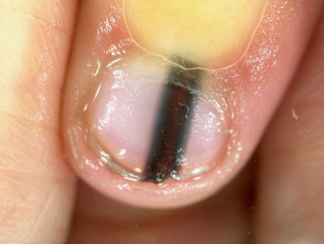
A female patient aged 60+ came into our podiatry clinic with a black longitudinal toenail line. This vertical dark streak running through her left big toenail was a cause of concern. A streak like this can be a sign of malignancy; a subungual melanoma. They are not to be ignored. Podiatrists are knowledgeable in nail pathologies. The treating podiatrist in this case was keen to know the history of this pigmentation. Fortunately, the patient recalled dropping an object on her toe, so it was diagnosed as a benign splinter haemorrhage. If she did not have a reason for the line’s appearance it could have been a different case, but the podiatrist was best placed to consider appropriate course of action. He would have referred the patient directly to her GP.
Differential Diagnosis
It can be very hard to distinguish a splinter haemorrhage from a melanoma. History and appearance of the longitudinal line are important determining factors for an experienced podiatrist to consider in an assessment. The line must not be confused with normal pigmentation, melanonychia ( increased pigmentation), found in darker skin type individuals. These pigmented lines can be divided into melanocytic and non-melanocytic. They are caused by increased melanocytic activation which results in an increase and depositing of melanin in new developing nail cells.
Melanonychia can be categorised as neoplastic, infective, systemic and drug-induced. Following examples:
- Neoplastic – Tumours
- Infective – Fungal nail infection, bacterial pseudomonas, verrucae, warts HPV
- Systemic – Renal disease, HIV, lupus, endocrine disorders
- Drug-Induced – Blood thinning drugs e.g. warfarin, chemotherapy drugs e.g. tetraycline can induce bleeding into nail plates.
Splinter Haemorrhage
Most splinter haemorrhages are caused by damage to nail bed/ plate structures, whether external or intrinsic. Small blood vessels are damaged underneath the nail plate. Classic characteristics are thin streaks which are black or dark brown in colour. When pressed they don’t disappear. As the nail plate growths forward, they grow out. Several nails can be affected.
Subungual Melanoma
Subungual melanomas are a form of skin cancer. Subungual melanomas fortunately are rare, accounting for only 1-3% of all diagnosed melanomas. 60% are found in fingernails and 40% in toenails. Subungual melanomas are more common aged 50+ and there can be a personal and family history of cancerous skin growths. They look very similar in colour and appearance to a splinter haemorrhage, but do not grow out with the nail plate and get wider in size. Usually larger than 3mm. A nodule can be noted under the nail and darkening of the skin at the beginning of the line into the cuticle. This is called the Hutchinson’s Sign. Only 1 nail is affected.
Subungual melanomas are rare and they can be hard to diagnose. If the clinician has any doubts then the patient must be urgently referred to their GP. The GP will in turn request a biopsy diagnostic examination. If the biopsy comes back positive for malignancy then a wide local excision of the melanoma is performed. The nail plate is removed, and the cancerous tissue excised. This can be done under local analgesia. Depending on how much tissue is excised the nail will grow out, but there may be some slight irregularity of the plate.
Importance of Removal
If a subungual melanoma is not detected or removed it could metastasize, spread to other parts of the body. As it is a cancer it is potentially life threatening, hence if you have no idea why you have a strange black line running through your nail plate, please get it checked out. Podiatrists are best placed to detect suspicious lesions like this.