
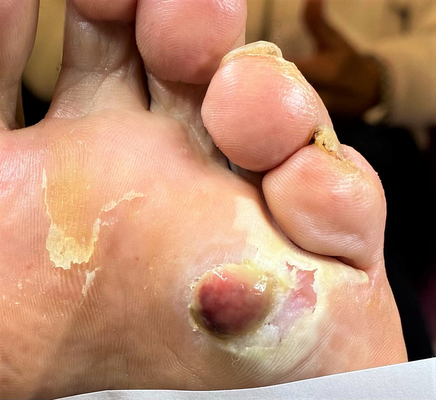
Last week I had a 74 year old female patient booked in with “ a painful hole in foot” resulting from home treating a verruca. As a podiatrist I was expecting to see a possible tissue breakdown or ulceration. Neither, something far more interesting and uncommon: pyogenic granuloma. These are not commonly seen on the sole of the foot.
Presentation
Presenting on the plantar sole aspect of her left foot was a small vascular looking, fleshy raised skin growth. This was directly where the verruca was situated. It was approximately 1.4 cm in size. Extremely tender on touch and bled easily at the base. It was attached by a ‘stalk’ to the base. Base was mascerated. No visible signs of any verruca tissue. The patient observed that it had a malodorous smell.
History
The patient had used a proprietary home freezing kit to freeze the verruca 5 weeks before her appointment. She froze the verruca twice in an attempt to eradicate it. No blister developed. This would have been the normal response to the cryotherapy. Instead, the fleshy tissue growth, grew quickly afterwards. This over-healing, granulation was caused as a direct response to the aggressive cryotherapy.
What is a Pyogenic Granuloma?
A pyogenic granuloma is a benign tumour. They mainly develop in younger ages groups and are common in pregnant women. Common places to occur are hands, fingers, arms, face, inside mouth and neck. It is uncommon to get them on the foot. They can occur as a direct response to trauma e.g. insect bite, hormones, drug interaction and infection. There is some suggestion that there is a viral cause.
In my patient’s case there was a direct link to confirm that the granuloma was caused by trauma. The quick onset also confirmed the diagnosis. Another factor which helped to confirm diagnosis, was my patient’s history of keloid scarring. She tended to develop pronounced raised scarring after trauma to the skin. She has compromised healing to skin trauma.
Why did it smell?
The unpleasant smell was bacterial. Staphylococcus Aureus can be found be found at pyogenic granuloma sites. It can be a causative factor. In my patient’s case there was considerable surrounding skin maceration which could be harboring the bacteria.
Foot Hypergranulation Tissue
Pyogenic granulomas are similar to hypergranulation tissue found in feet. Both are forms of over-granulation. Patients with chronic ingrown toenails present with proud fleshy tissue, commonly around the area of concern. Hypergranulation tissue like a pyogenic granuloma is very vascular and bleeds readily on touch.
Hypergranulation is basically an over-healing response, whereby the normal process of tissue granulation to form new connective tissue and tiny blood vessels goes into over-drive. An over-proliferation of healing tissue. Unlike a pyogenic granuloma, hypergranulation is more irregular in development and can be expansive. There is no stalk/ stem base.

Treatment Pyogenic Granuloma
My patient was limping, the granuloma was very painful on weight-bearing. It need removing and for normal healing to be mediated. After application of LMX4 cream and injecting with 3% plain Mepvicaine, the granuloma was excised at the base. Silver Nitrate was applied to cauterize the base. An Inadine dressing was applied and deflective padding.
The patient was reviewed 5 days later. She came into clinic without a limp and was delighted that the discomfort had dissipated. After debriding the silver nitrate eschar, there was nice new pink skin underneath and no maceration. Another application of silver nitrate was applied directly onto the centre of the wound. This should be enough to ensure any over- granulating tissue shrinks back.
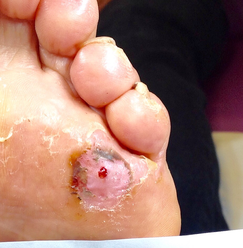
On a good positive note there was no sign of any verruca!